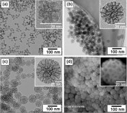
Human colon carcinoma cells (HT29) chosen as a model system for the study of the uptake of Ptnanoparticles. In order to control the particle properties, Pt nanoparticles are produced on β-cyclodextrin (β-CD) by supercritical fluid reactive deposition (SFRD) in the group of M. Türk (Institut für Technische Thermodynamik und Kältetechnik). Depending on the organometallic precursors and process conditions, SFRD enables the deposition of pure and highly dispersed Pt nanoparticles with distinctly different sizes and shapes. This is illustrated in Figs. 1a-c, showing as examples a single decahedral nanoparticle with about 40 nm size, an agglomerate of very small (~5 nm) irregularly-shaped particles, and approximately 100 nm sized Pt particles with predominantly cuboctahedral shape. The cellular uptake and biological response is studied in dependence on the mean particle size, particle concentration, and time of incubation in the group of D. Marko (Institut für Angewandte Biowissenschaften, Abteilung für Lebensmittelchemie und Lebensmitteltoxikologie). A significant damage of the DNA is observed for small nanoparticles with sizes below 30 nm.
Pt nanoparticles in the cells can be directly visualized by using scanning electron microscopy (SEM) and focused ion-beam (FIB) milling HT29 cells. The cells are grown on Transwell membranes and prepared for the SEM/FIB studies by fixation, dehydration and critical-point drying. Fig. 2a shows a secondary-electron (SE) image of a single HT29 cell after 24 h Pt (1 μg/cm2) incubation. The surface of the HT29 cell is characterized by microvillis and some phagocytotic protrusions. In the backscattered-electron (BSE) image of the same cell (Fig. 2b) agglomerates of Pt particles are revealed on the cell surface (arrows). This HT29 cell was subsequently cut “slice-by-slice” with the focused Ga+ ion beam in the FIB instrument and imaged by BSE. In this way, Pt particles were found in different depths in the cell interior (cf. Fig. 2c) demonstrating cellular uptake. Moreover, as visible in Fig. 2d the presence of platinum was detected by energy-dispersive X-ray spectroscopy (EDXS).
Selected publications:
[1] J. Pelka, H. Gehrke, M. Esselen, M. Türk, M. Crone, S. Bräse, T. Muller, H. Blank, W. Send, V. Zibat, P. Brenner, R. Schneider, D. Gerthsen, D. Marko, Cellular Uptake of Platinum Nanoparticles in Human Colon Carcinoma Cells and Their Impact on Cellular Redox Systems and DNA Integrity,
See also:- Georgiaapostille.us - Hague Apostille Member Countries - Georgia apostille, Atlanta apostille.


 Because I believe
  One who loves God will see what they believe and the ones who trust science will believe what they see


